Art
Knockdown of the Rp-dpp gene from Rhodnius prolixus by RNA interference
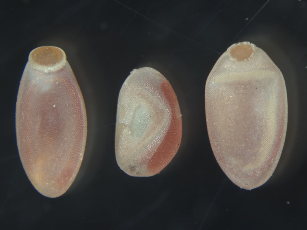
The chorion surrounding the embryos subjected to Rp-dpp RNAi frequently shows absence of the operculum and deformities.
Drosophila melanogaster embryos at the blastoderm stage
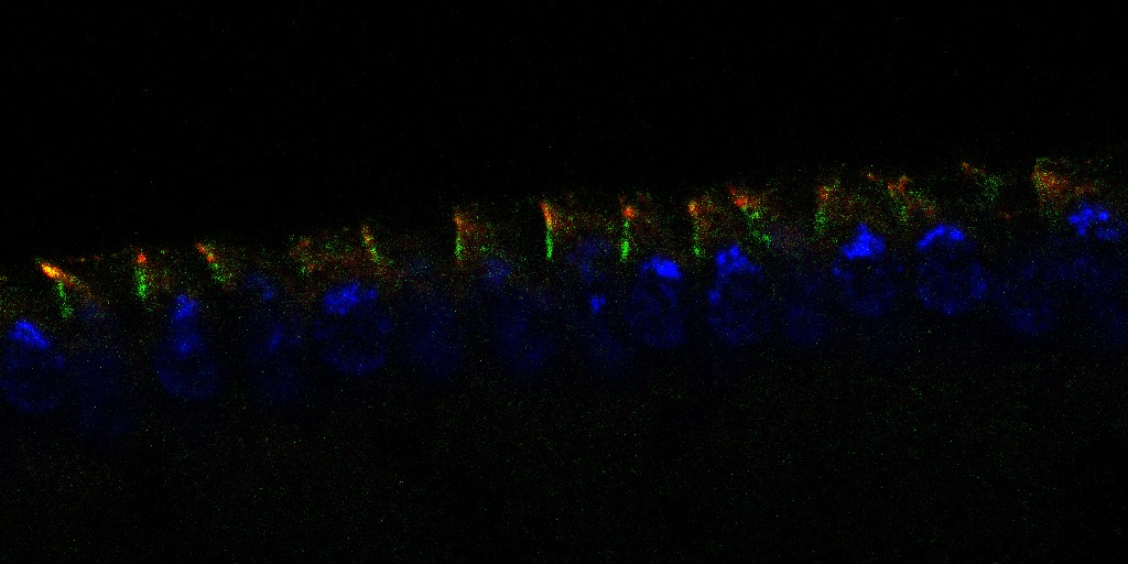
Embryos labeled with antibodies against the Toll receptor and Peanut protein, which marks the plasma membrane during the cellularization process. Images acquired using confocal microscopy.
The Bone Morphogenetic Protein (BMP) signaling pathway
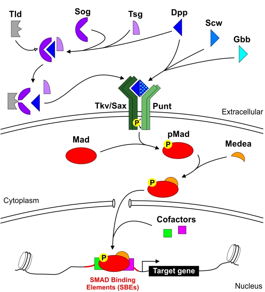
Schematic illustration of the BMP pathway in Drosophila melanogaster, showing receptor activation, Smad-mediated signaling, and its role in gene regulation.
Detection of Rp-engrailed by in situ hybridization in Rhodnius prolixus embryo
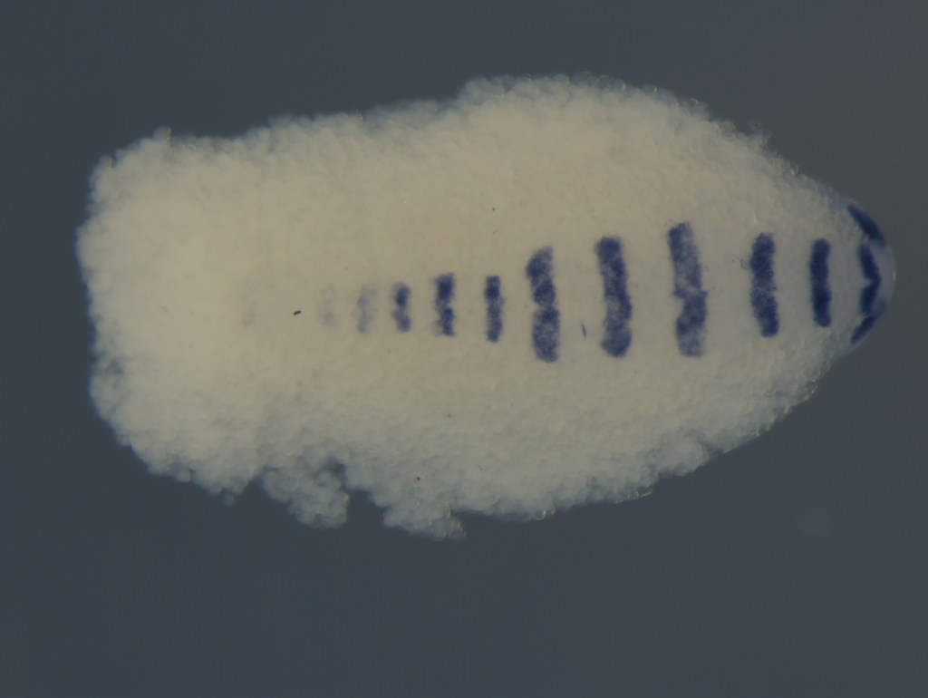
Expression of the engrailed gene in a stage 4 embryo, revealed by in situ hybridization, highlighting the spatial localization of the transcript during embryonic development.
Histological section of Rhodnius prolixus embryo during gastrulation
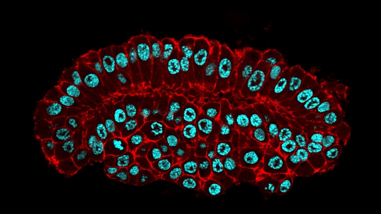
Germ band of the R. prolixus embryo labeled with phalloidin (red) for filamentous actin and nuclei (blue) of the mesoderm, ectoderm, and amnion.
Muscles of Drosophila melanogaster larva
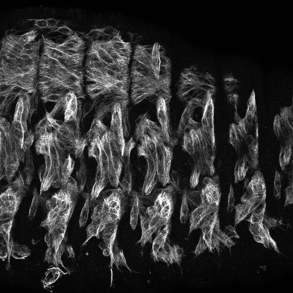
The repetitive muscle pattern in each body segment, revealed by phalloidin staining (filamentous actin).
Eye of the kissing bug Rhodnius prolixus
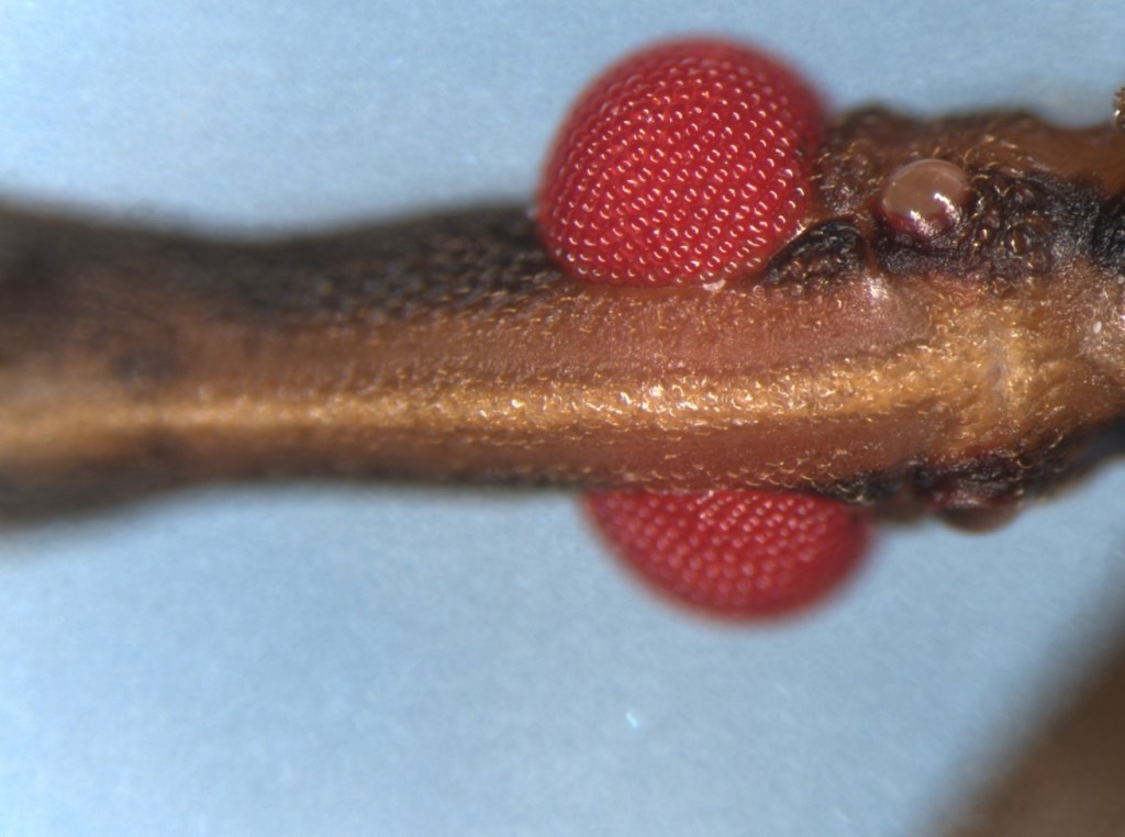
Spontaneous mutant of R. prolixus showing a red eye instead of the typical black.
Ovarian follicle of Drosophila melanogaster with mutant cell clones for the sog gene
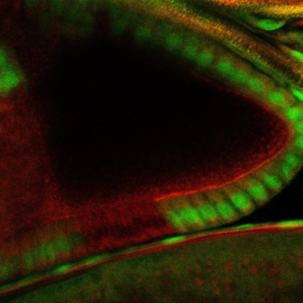
The image identifies mutant cells lacking sog expression, marked by the absence of GFP (green). Mutant cell clones were generated using the FLP/FRT system.
Address
Laboratório de Biologia Molecular do Desenvolvimento – Centro de Ciências da Saúde (CCS), Bloco F, sala F2-031 – UFRJ – Cidade Universitária – Rio de Janeiro – RJ – Brasil ZIP 21941-902
Designed with WordPress
